
All PIP Joint Flexion Contractures Are Not the Same

March 2025 No. 89
All PIP Joint Flexion Contractures Are Not the Same
by Judy Colditz, OT/L, CHT, FAOTA
There are many causes of proximal interphalangeal (PIP) joint flexion contractures in the fingers with two common causes being proximal phalanx fractures that cause adherence of the dorsal apparatus to the bone, and PIP joint dislocations that disrupt the capsule of the joint causing the surrounding structures to adhere during healing. We will look in detail at how these two injuries differ and thus how the approach to resolving the resultant PIP flexion contractions should be modified to focus on the exact location of tissue adherence.
Anatomy of a Proximal Phalanx Fracture
The thin but expansive dorsal apparatus (DA), (See Figure 1) comprises multiple fibers running in a variety of directions and lies directly over the proximal phalanx, in some locations covering as much as half of its surface. See Figure 2. It is not just the proximal and distal glide of the dorsal apparatus that creates interphalangeal joint extension and allows finger flexion, but it is also the change of angle of the fiber layers relative to one another that allow it to “expand” and “contract.”

Figure 1: A cadaveric specimen of the dorsal apparatus of the finger.
With no intervening tissue layer, the dorsal apparatus sits directly on the bone. See Figure 2. Therefore, the matrix formed from the bleeding proximal phalanx fracture adheres the dorsal apparatus fibers to the healing bone. This adherence requires us to focus on early motion of proximal phalanx fractures because adherence of the dorsal apparatus to the proximal phalanx limits full finger extension and may limit full finger flexion.

Figure 2: Cross-section at the level of the proximal phalanx showing the thin dorsal apparatus over the proximal phalanx; Cropped image with permission from Meals & Seeger: "An Atlas of Forearm and Hand Cross-Sectional Anatomy", 1991.
Anatomy of a Proximal Phalanx Dislocation
Most commonly the PIP joint is dislocated dorsally. In other words, the middle phalanx moves dorsal in relation to the proximal phalanx. See Figure 3. A dorsal dislocation of the PIP joint injures the volar plate and disrupts the joint capsule.

Figure 3: X-ray of dorsal PIP joint dislocation.
At the level of the PIP joint the lateral edges of the DA (the lateral bands) are more lateral than over the proximal phalanx. When the PIP joint actively flexes, they move further laterally and then volarly. See Figure 4. This means they must then move dorsally to assist with active PIP joint extension. Whereas proximal glide of the DA over the proximal phalanx is required to achieve PIP joint extension, at the PIP joint itself, there must be a combination of proximal glide of the dorsal apparatus (the central slip being the most dorsal structure) as well as dorsal and proximal glide of the lateral structures (the lateral bands).

Figure 4: Dorsal view of cadaver finger shows how the lateral bands sit laterally at the PIP joint.
Keep in mind, in the example of the PIP joint dorsal dislocation, the proximal glide of the DA is not impeded over the proximal phalanx. But the central slip insertion cannot alone extend the PIP joint. The moment arm [the perpendicular distance between the joint and a line of force] of the central slip insertion is short, making it a relatively inefficient extensor of the PIP joint. For PIP joint extension to occur, all the tendinous fibers around the PIP joint must be able to move in addition to the central slip and the proximal fibers of the dorsal apparatus.
During normal finger flexion, the lateral bands at the PIP joint move from a lateral position to a more volar one although some would argue at the extreme, they move below the axis. But in the normal finger the intact fibers of the dorsal apparatus pull the lateral bands dorsally as the finger extends. The more dorsal the position of the lateral bands, the more they participate in PIP joint extension.
The inefficiency of the lateral bands when the PIP joint is in flexion explains why it is useful to employ some type of orthotic intervention until the PIP joint flexion contracture is approximately -45°. It is only at this approximate angle that the lateral bands can begin to effectively participate! Therefore, following a PIP dorsal dislocation, often the structure that needs to move the most at the PIP joint during finger extension is the lateral bands.
It is useful to remember that the lateral bands are nothing more than the thickened edge of the dorsal apparatus, they are not distinctly separate tendinous structures. See Figure 1. Therefore, during normal finger flexion and extension, not only must the dorsal apparatus move and change configuration over and around the PIP joint, but the lateral bands must also move dorsally during extension, and laterally/volarly during flexion.
PIP Joint Flexion Contractures
Individuals with long-standing ulnar palsy who have developed resistive PIP joint flexion contractures of the ring and little fingers can resolve these contractures with active motion only when the metacarpophalangeal (MP) joint is blocked in flexion. See Figure 5. This is proof that active motion can resolve a passive deficient.
Therefore, the author has abandoned previously popular approaches of serial casting and other orthotic fabrication to remedy PIP joint flexion contractures unless the contracture is severe. In that case, serial casting is needed to bring the contracture to less than 45° of flexion so the lateral bands can actively participate in PIP joint extension.
The author is adamantly opposed to passive range of motion or dynamic splinting to achieve PIP joint extension simply because these approaches do not recruit the muscle pull on the fibers that must move to create the motion.


Figure 5: Ulnar palsy created clawing of the ring and little fingers which leads to PIP joint flexion contractures; this can be prevented or resolved by blocking MP joint hyperextension with an orthosis.
WHAT IS THE DIFFERENT TREATMENT FOR PIP JOINT FLEXION CONTRACTURES CAUSED BY A PROXIMAL PHALAX FRACTURE VERSUS A PIP JOINT DORSAL DISLOCATION?
Treatment Following Proximal Phalanx Fracture
Following a proximal phalanx fracture the single most important focus is to assure movement of the dorsal apparatus over the proximal phalanx. One commonly sees hyperextension of the metacarpophalangeal joint with attempted active extension of the PIP joint. See Figure 6. This MP joint movement uses up the proximal excursion of extensor digitorum communis (EDC) making it less available to contribute to interphalangeal joint extension.
Therefore, assuming the reader is approaching PIP flexion contractures with active motion, the obvious choice is to block MP joint extension to redirect the power of the EDC into the dorsal apparatus via the central slip and ultimately the central slip insertion. Since the central slip inserts just distal to the PIP joint there is relatively little excursion at this level. But the proximal excursion of the DA is essential to achieve full PIP joint extension.

Figure 6: Active finger extension when a PIP joint flexion contracture is present results in MP joint hyperextension.
The angle at which the MP joint should be blocked depends on the severity of the PIP joint flexion contracture as well as the opposing need for regaining finger flexion. A useful guideline is the greater the PIP joint flexion contracture, the more the MP joint should be blocked in flexion.
This principle can be carried out with a multitude of designs. See online lecture: “The Obstinate PIP Joint” for a video demonstration of three orthoses. One current popular design is a relative motion orthosis, blocking the MP of the injured finger in relatively more flexion than the adjacent MP joints. See Figure 7.

Figure 7: Relative motion orthosis blocks extension of the ring finger MP joint, driving all extension forces to the PIP joint.
But if only one finger is involved, a relative motion orthosis is restrictive for keyboarding. For keyboarding, not only is there an advantage of including only the MP joint of the injured finger, but it is helpful for the patient to allow greater MP joint extension when on the keyboard while still preventing MP joint hyperextension. In such patients it may be helpful to provide something simple that can be adjusted to block the MP joint at the ideal angle while still allowing effective typing. See Figures 8 & 9.

Figure 8: This orthosis, made of leather, hook & loop, and string allows adjustment of the MP joint position.

Figure 9: Adjusting the MP joint extension block allows normal typing postures while still preventing MP joint hyperextension.
Treatment Following a PIP Joint Dislocation
Because the extensor digitorum communis is an extrinsic muscle with greater power than the intrinsic finger muscles it is always useful to harness its energy actively toward PIP joint extension by blocking the MP joint. But in the case of a PIP joint dislocation where the healing tissue is more specific to the joint itself, it may be helpful to also increase the tension on the lateral bands to assist in their required dorsal/volar movement at the PIP joint.
After the lateral bands cross the PIP joint, they begin to move dorsally on each side of the middle phalanx, before coming together immediately prior to the terminal tendon insertion on the dorsum of the distal interphalangeal (DIP) joint. Therefore, passively flexing the DIP joint increases tension on the lateral bands. Increased tension allows the lateral bands to improve its participation in PIP joint extension. See Figure 10.


Figure 10: Top photo shows active PIP joint extension without orthosis and bottom shows improvement in active PIP extension with DIP joint blocking orthosis. NOTE: Bulbous dorsum of PIP joint makes it appear more flexed.
Not only is blocking the DIP joint in flexion useful for patients following a PIP joint dislocation but also following a boutonniere injury when a PIP joint extension lag is present. A simple DIP joint blocking orthosis can be made of molded thermoplastic material or a piece of aluminum with foam padding. The orthosis is taped circumferentially, allowing further DIP joint flexion but blocking full extension. See Figure 11.
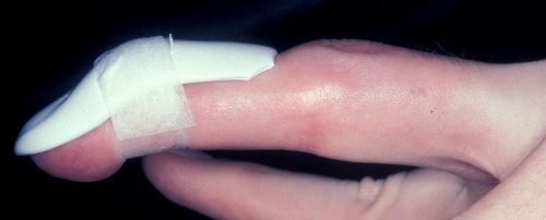
Figure 11: A small piece of molded thermoplastic material taped on the dorsum of the finger can serve as a DIP joint extension block.
It is logical to begin with a position of greater DIP joint flexion but one must be cautious not to overdo, which can create an extension lag at the DIP joint. As the patient improves PIP joint extension, the angle of the DIP flexion block decreases and then weaning occurs.
Looking Differently
This description of a diverse approach to two differing causes of PIP joint flexion contractures is a simple overview. The biomechanics of PIP joint extension are far more complex than can be covered in this brief explanation. However, we hope this explanation allows you to differentiate when just blocking the MP joint is adequate and when perhaps adding a DIP extension block assists in regaining better active PIP joint extension.
The reader will note that exact angles of MP joint blocking have not been specified. Because every patient is different and clinical judgment must determine what is most advantageous for each individual patient, an exact protocol does not exist. Suffice it to say that the greater the PIP flexion contracture the greater the position of the MP joint flexion should be. That statement can also be applied to blocking the DIP joint to improve lateral band excursion at the PIP joint but with a much greater caution to avoid creating a mallet finger. One must, however, always consider the opposing need for regaining flexion. Although finger flexion is stronger than extension, if the flexor system has been injured, the above statement about the MP joint position must be modified.
If the reader is interested in learning more about the PIP joint, the following online resources are recommended:
1. Drawing the Dorsal Apparatus
2. Understanding the Dorsal Apparatus of the Finger
3. What Drives Finger Motion
4. Insight into the Interosseous Muscles
5. The Dorsal Extensor Apparatus - Simplified and Discussed (GK Hand Surgery)
Download Clinical Pearl No. 89, All PIP Joint Flexion Contractions Are Not the Same, March 2025
© BraceLab; 2025 all rights reserved
Disclaimer: BraceLab Clinical Pearls are intended to be an informal sharing of practical clinical ideas; not formal evidence-based conclusions of fact.