AI Gait Analysis Reveals the Unseen Impact of an Ankle Brace

August 2026 No. 49
AI Gait Analysis Reveals the Unseen Impact of an Ankle Brace
See how the AI Mov-Scan objectively measured functional changes with the Push® med Ankle Brace Aequi Flex
by Jerry Ditz, DPT, Dip. Osteopractic, Cert. SMT, Cert. DN
Clinical Problem
Ankle sprains are typically treated as simple injuries. A patient injures the ankle, manages swelling, wears a brace, and eventually returns to daily life. In many cases, pain improves and the patient returns to normal activity. As clinicians, we know that symptom improvement does not always mean fully normalized movement.
Many patients continue with a compensatory gait which is usually subtle and, thus, difficult to detect during a clinical exam. Gait variations become more apparent when the patient progresses from walking to running.
Finding The Answers
BraceLab evaluated Baliston Health's AI Mov-Scan (Figure 1) to objectively measure how the Push® med Ankle Brace Aequi Flex (Figure 2) influences movement during recovery from an ankle sprain.
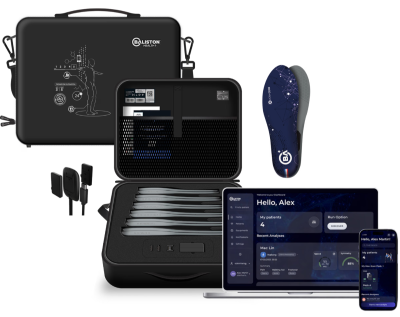
Figure 1: Baliston Health's AI Mov-Scan kit
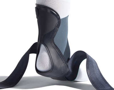
Figure 2: Push® med Ankle Brace Aequi Flex
Rather than relying solely on symptom improvement or clinical observation, we wanted to quantify how gait changed with and without the brace. Using AI gait analysis, we evaluated walking and running mechanics throughout recovery to better understand the brace's functional impact.
While this single-patient case does not establish product efficacy, it demonstrates how AI gait analysis can provide objective insights into patient movement.
In this case we wanted to obtain answers to specific questions. When wearing the brace:
• Does the patient load the injured side more evenly?
• Does walking symmetry improve?
• Does push-off become more efficient?
• Is excessive supination reduced?
• When the patient progresses to running, are there still deficits that walking does not reveal?
Case Example
A 41-year-old male fitted with the Aequi Flex ankle brace one day after a grade II right ankle sprain was evaluated with the AI Mov-Scan. The following evaluations were completed:
INITIAL: One day post injury
1. Walking without the brace
2. Walking immediately after applying the brace
3. Walking after wearing the brace for approximately one hour
FOLLOW-UP: 10 days post injury
1. Walking without the brace
2. Walking with the brace
3. Running without the brace
4. Running with the brace
Walking Evaluations
After wearing the ankle brace for 9 days, the patient's ankle pain improved from 4/10 to 1/10. Although the pain improvement was clinically meaningful, the objective gait evaluations better described the changes occuring throughout the recovery process.
Early in recovery, the Aequi Flex ankle brace improved walking symmetry, contact time balance, and overall gait quality after a brief acclimation period.
As pain subsided and gait normalized, the brace's influence became more specific. Rather than further improving overall gait scores, it helped reduce right-side supination while preserving normal walking mechanics.
Walking With the Brace During Early Recovery: Improved Symmetry
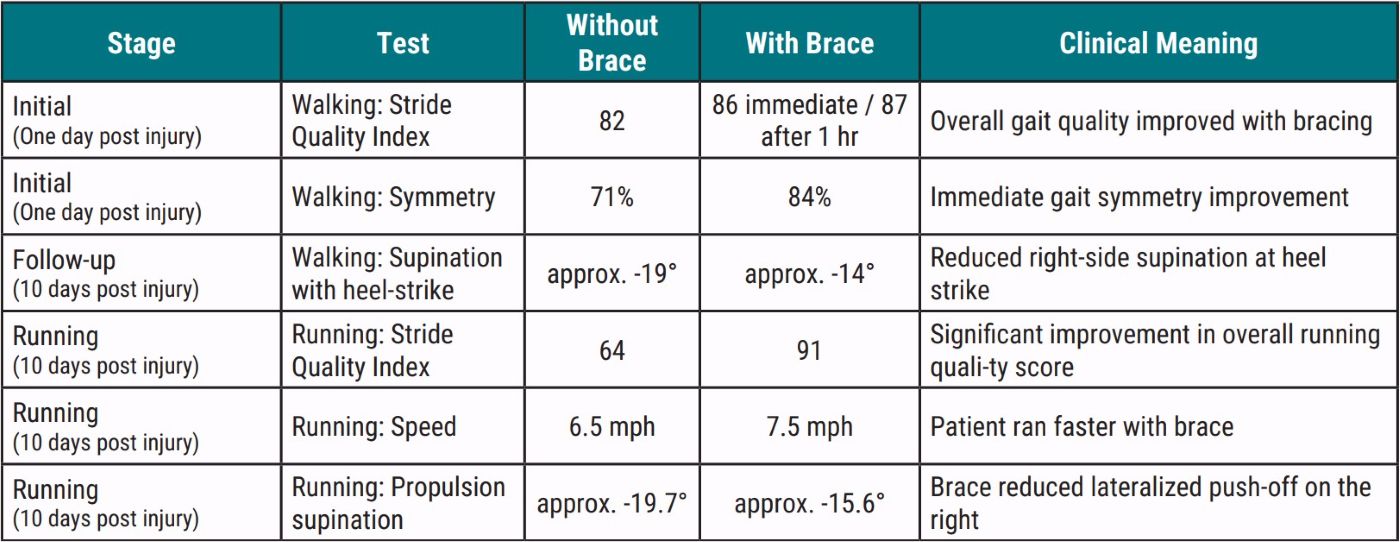
At the initial walking test, the patient demonstrated a protective gait pattern and reported right ankle pain of 4/10. With brace wear, the Stride Quality Index* (SQI) increased from 82 without the brace to 86 immediately after application and to 87 after one hour. Because the SQI is a composite measure of gait symmetry, pronation control, propulsion efficiency, shock absorption, variability, and selected risk factors, this improvement suggests a modest overall shift toward a more efficient, stable, and symmetrical gait pattern. The symmetry-specific measures showed a more pronounced change: global symmetry improved from 71% to 84%, while walk-profile asymmetry decreased from 7.6% without the brace to 2.5% immediately with the brace and to 0.7% after one hour.
*Stride Quality Index is an overall walking quality score based on key gait parameters such as stride time, stance/swing time, double support time, stride length, foot progression angle, pronation/supination angle, and ankle position at gait phases. The higher the score the closer a gait pattern is to typical norms.
Sample Reports
For the reader to better appreciate the information provided by Baliston Health's AI Mov-Scan, data reports below have been excerpted from the Mov-Scan App. You may see the full reports for all tests here:
• Walk Test: Sprained Ankle (no brace)
• Walk Test: Sprained Ankle (w/ brace)
• Walk Test: Sprained Ankle (wearing brace for 1 hour)
• Walk Test: Sprained Ankle (1 week later, no brace)
• Walk Test: Sprained Ankle (1 week later, w/brace)
Mov-Scan Walk Test: Sprained Ankle
AI Mov-Scan Interpretation (See Graph 1)
INITIAL EVALUATION: Walking without the brace
Walk Profile AI Interpretations:
• Your patient exhibits a mild but clinically meaningful asymmetry in stance time (left > right: 7.6%), with the left side spending 26 ms longer in stance than the right. This suggests a subtle compensatory shift in load distribution, likely to offload the right ankle given the reported pain (VAS 4/10). The prolonged left stance time (63.2% of gait cycle vs. 60.8% right) and reduced right swing time proportion (39.2% vs. 36.8% left) further support a right-side avoidance strategy during propulsion. While stride length remains symmetrical (1.4 m both sides), the prolonged stance phase on the left may indicate reduced push-off efficiency on the right, potentially increasing cumulative stress on the left limb over time. The double support time proportion (12%) is within normal limits, suggesting no significant balance impairment. These findings align with a protective adaptation to right ankle discomfort, warranting targeted assessment of right ankle mechanics (e.g., talocrural joint mobility, peroneal strength) and potential gait retraining to restore symmetrical propulsion.
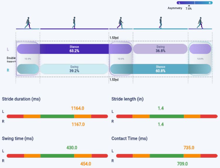
Graph 1: Walk test - Walking without the brace - Walk Profile
AI Mov-Scan Interpretation (See Graph 2)
INITIAL EVALUATION: Walking without the brace
Supination/Pronation Angles:
• Provides the angles of pronation/supination during the 4 key stages of the contact phase (heel strike, toes contact, heel off, toes off).
• The graph shows the average values (colored lines) as well as the standard deviations (thin lines), and the statistical norms (green corridor).
Pronation/Supination Angles AI Interpretations:
• Your patient demonstrates consistently high supination angles across all gait phases (heel impact, foot flat, and propulsion), with the right side exhibiting more pronounced deviations (e.g., -21.3° at heel impact vs. -15.7° left).
• This excessive supination, particularly on the right, may reflect residual biomechanical adaptations following the reported ankle sprain, potentially increasing lateral ankle strain and predisposing to reinjury or chronic instability.
• The asymmetry (right > left) aligns with the patient's reported right ankle pain (4/10), suggesting a localized dysfunction rather than a systemic issue. While no stride or timing abnormalities were flagged, the supination pattern alone warrants targeted proprioceptive or strengthening interventions to restore neutral foot positioning during gait.
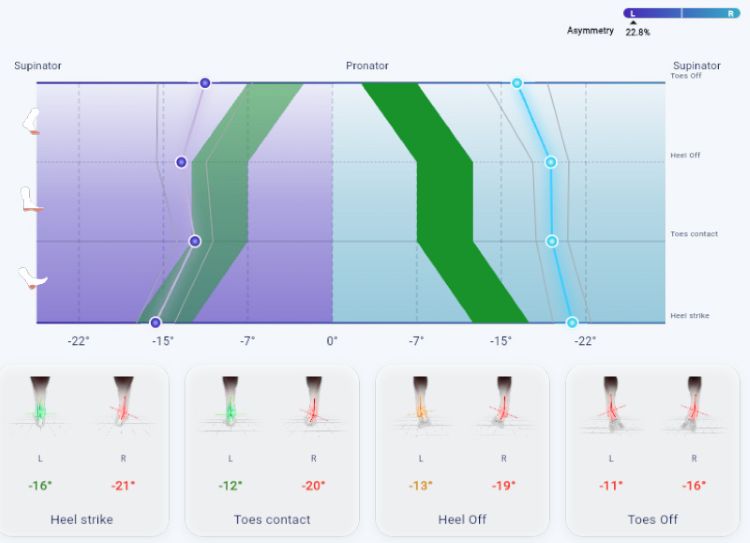
Graph 2: Walk test - Walking without the brace - Supination/Pronation angles
Mov-Scan Walk Test: Sprained Ankle (w/ brace)
AI Mov-Scan Interpretation (See Graph 3)
INITIAL EVALUATION: Walking immediately with the brace
Walk Profile:
• Average duration of the stance and swing phases related to the total cycle time.
• Normal: stance 60% and swing 40%
• In case of an asymmetry, there is a compensation by the stronger limb (longer stance time), and potential limping.
Walk Profile AI Interpretations:
• Your patient presents with prolonged stance and swing phases bilaterally, indicating a generalized reduction in gait cadence (stride time ~1.17 s).
• While stance and swing times are symmetrically prolonged, the absence of meaningful asymmetry suggests a compensatory adaptation rather than a unilateral deficit, likely influenced by the reported right ankle pain and use of orthopaedic shoes.
• The average stride length (1.4 m) and double support time fall within normal ranges, but the combined increase in stance/swing duration implies reduced propulsion efficiency, which may contribute to fatigue or overuse strain in the lower limbs.
• The pain intensity of 4/10 could further exacerbate these deviations by encouraging a protective gait pattern, though no severe asymmetry is detected.
• Clinically, this pattern warrants focused assessment of ankle joint mobility and footwear efficacy, particularly given the history of ankle sprain and orthotic use.
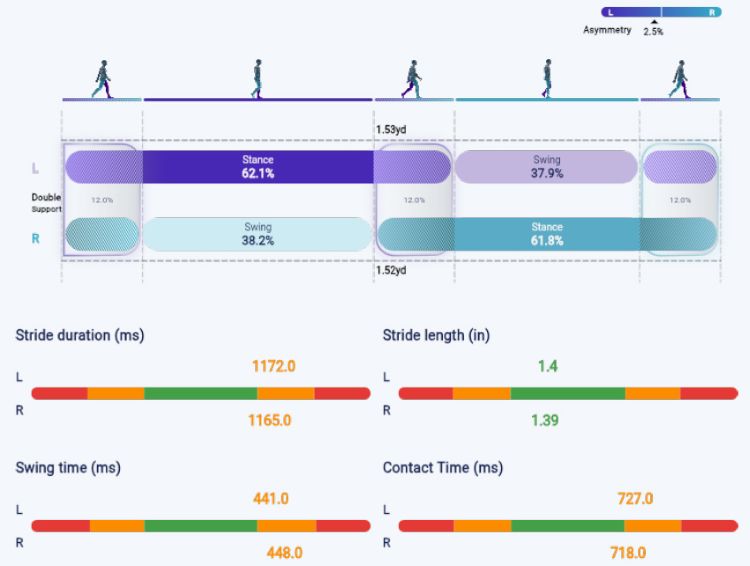
Graph 3: Walk test - Walking immediately with the brace - Walk Profile
AI Mov-Scan Interpretation (See Graph 4)
INITIAL EVALUATION: Walking without the brace
Supination/Pronation Angles:
• Provides the angles of pronation/supination during the 4 key stages of the contact phase (heel strike, toes contact, heel off, toes off).
• The graph shows the average values (colored lines) as well as the standard deviations (thin lines), and the statistical norms (green corridor).
Pronation/Supination Angles AI Interpretations:
• Your patient exhibits consistent supination dominance throughout the gait cycle, with right-side supination angles exceeding clinically normal ranges at all phases (heel impact, foot flat, and propulsion).
• This pattern suggests a compensatory supination strategy, likely linked to prior ankle sprain history and the use of orthopedic shoes, which may reduce medial foot-ground contact and shift load laterally.
• The right ankle's supination angles (-18.9° at heel impact, -18.5° at foot flat, -13.7° at propulsion) are markedly elevated compared to the left, indicating asymmetric foot positioning that could predispose to lateral ankle strain, reduced shock absorption, or altered knee/hip mechanics.
• Given the reported pain intensity (4/10) in the right ankle, this supination bias may exacerbate joint stress during propulsion.
• The findings warrant targeted strengthening of peroneal and tibialis posterior muscles to counteract supination torque, alongside gait retraining to encourage controlled foot pronation during stance.
• Orthopedic shoe use may need adjustment to ensure proper medial arch support.
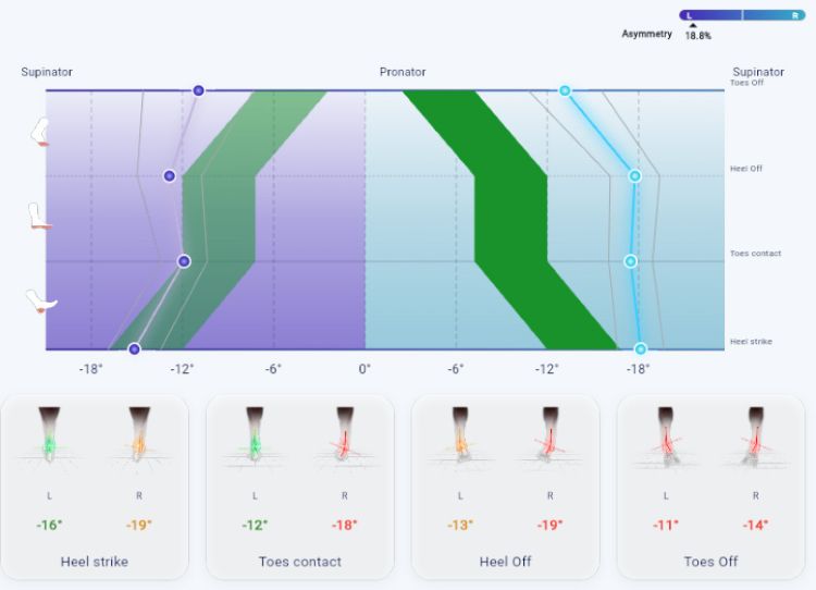
Graph 4: Walk test - Walking immediately with the brace - Supination/Pronation angles
Mov-Scan Walk Test: Sprained Ankle (w/ brace wearing brace for 1 hour)
AI Mov-Scan Interpretation (See Graph 5)
INITIAL EVALUATION: Walking after 1 hour in brace
Walk Profile:
• Average duration of the stance and swing phases related to the total cycle time.
• Normal: stance 60% and swing 40%
• In case of an asymmetry, there is a compensation by the stronger limb (longer stance time), and potential limping.
Walk Profile AI Interpretations:
• The gait analysis reveals a normal temporal-spatial pattern with no clinically meaningful deviations in stride length, swing/stance proportions, or symmetry.
• The 11.4% double support time proportion is within normal limits, suggesting no compensatory instability or cautious gait pattern typically seen in acute ankle pathology.
• The right ankle pain (4/10) may contribute to subtle adjustments, but the absence of asymmetry in stance time (0.7% difference) and balanced swing/stance phases indicates no significant biomechanical compensation at this stage.
• The findings suggest mild functional adaptation rather than structural dysfunction, though further clinical correlation with the reported ankle sprain is warranted to assess residual impairment.
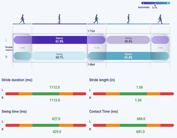
Graph 5: Walk test - Walking with the brace after wearing brace for 1hr - Walk Profile
AI Mov-Scan Interpretation (See Graph 6)
INITIAL EVALUATION: Walking without the brace
Supination/Pronation Angles:
• Provides the angles of pronation/supination during the 4 key stages of the contact phase (heel strike, toes contact, heel off, toes off).
• The graph shows the average values (colored lines) as well as the standard deviations (thin lines), and the statistical norms (green corridor). In case of an asymmetry, there is a compensation by the stronger limb (longer stance time), and potential limping.
Pronation/Supination Angles AI Interpretations:
Your patient exhibits a consistent supination-dominant foot posture throughout the gait cycle, with the right side demonstrating more pronounced deviations than the left. At heel impact, propulsion, and foot flat phases, the right foot shows high supination (up to -19.3°), while the left remains within neutral to mild supination ranges. This pattern suggests a rigid foot mechanics during weight acceptance and push-off, which may reduce shock absorption efficiency and increase repetitive strain on the lateral ankle structures — particularly relevant given the history of right ankle sprain. The asymmetry in supination angles (right > left) could indicate either a primary right foot dysfunction or a compensatory adaptation to prior injury. The neutral-to-mild supination on the left suggests it compensates by absorbing more load, potentially altering lower limb alignment. These findings may contribute to the reported right ankle pain (4/10) by increasing lateral joint stress during gait.
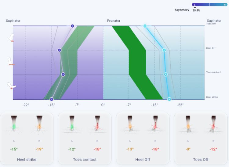
Graph 6: Walk test - Walking with the brace after wearing brace for 1hr - Supination/Pronation angles
Walking With the Brace During Follow Up Testing: The Brace Improved Supination
At follow-up testing (10 days post injury), while global walking scores with the brace had not improved further, ankle pain had decreased to 1/10 and walking without the brace was much improved. The brace, however, provided specific mechanical benefits:
• Right heel-strike supination improved from approximately -19° without brace to approximately -14° with the Aequi Flex ankle brace
• Right toe-off supination improved from approximately -12° to approximately -10°.
Excessive supination positions the foot toward inversion, the mechanism commonly associated with lateral ankle sprains, and may increase stress on the healing lateral ankle ligaments. The reductions observed with the brace suggest that it helped limit this position during both initial contact and push-off, even though the patient's overall walking scores had already improved as pain and natural recovery progressed.
Walking Changes Over Time
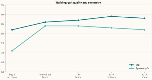
Figure 3: Walking SQI and symmetry improved early with bracing and stayed stronger at follow-up.
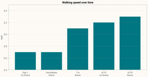
Figure 4: Walking speed improved over time; the fastest walking trial was at follow up with brace wear.
Running Evaluations
Sample Reports
For the reader to better appreciate the information provided by Baliston Health's AI Mov-Scan, data reports below have been excerpted from the Mov-Scan App. You may see the full reports for all tests here:
• Run Test: Sprained Ankle (1 week later, no brace)
• Run Test: Sprained Ankle (1 week later, w/brace)
Running With the Brace: Higher Demand Revealed Persistent Deficits
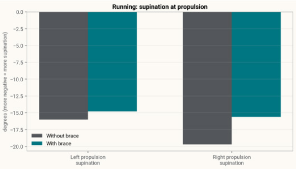
The initial evaluation, when the pain and compensation were obvious, was compared to a running evaluation during the follow up evaluation 10 days post injury. At the follow up evaluation, walking had improved, but running exposed deficits that were not obvious during walking. Without wearing the brace, running SQI was 64, cadence was 153 steps/min, rearfoot strike dominance persisted, propulsion speed was very low bilaterally, and right propulsion supination was approximately -19.7°. When wearing the Aequi Flex ankle brace, running SQI increased to 91, speed increased from 6.5 to 7.5 mph, cadence increased slightly to 157 steps/min, propulsion speed improved, and right propulsion supination improved to approximately -15.6°.
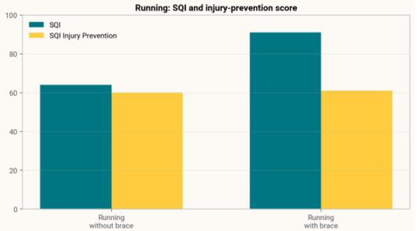
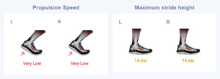
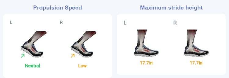
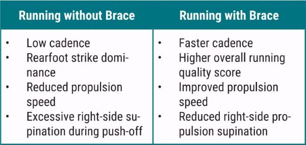
Although walking without the brace had significantly improved, running without the brace demonstrated continued dysfunction. Although improved, running mechanics did not fully normalize wearing the brace:
Combined Findings
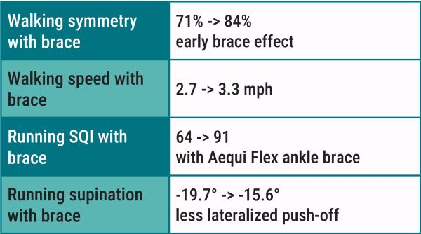
In this case example, Baliston Health's AI Mov-Scan associated the Aequi Flex ankle brace with measurable beneficial changes in walking and running mechanics after a right ankle sprain. Early walking tests showed improved symmetry and better functional walking after a short acclimation period. Later walking tests suggested that, even when pain was low and gait had improved, the brace helped reduce right-side supination while preserving normal walking mechanics. During running, the brace was associated with improved overall running quality, faster speed, improved propulsion speed, and reduced right-side propulsion supination.
A brace can be helpful, but it is not magic. Using AI gait analysis while wearing a brace moves the conversation from: "Does the brace feel supportive?" to "What changed in the way the patient moves?"
An effective brace can support better joint mechanics, improve confidence, and increase lateral ankle control, especially during higher-demand activity. In this case, it appears to support lateral ankle control during running, although it did not fully normalize running mechanics. Cadence remained low, rearfoot strike dominance persisted, right propulsion remained lower than left, and vertical stride height increased during the braced run.
Use of a brace must always be paired with appropriate rehabilitation for progressive strengthening, proprioceptive training, cadence retraining, and running-form work. This single-patient case example is not proof of product efficacy so one should interpret these findings accordingly.