
Boutonniere Basics

July 2026 No. 97
Boutonniere Basics
by Karol S. Young, OTD, OTR/L, CHT
When a patient presents with a stiff flexed proximal interphalangeal (PIP) joint and extended distal interphalangeal (DIP) joint, how do you know if it is a boutonniere (injury to the dorsal apparatus) or a joint flexion contracture (often called a pseudo-boutonniere) due to trauma to the PIP joint? (See Figure 1.)
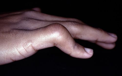
Figure 1: What is the diagnosis?
In absence of a definitive diagnosis, focusing on the anatomy can save you from frustration, misdiagnosis, and inappropriate orthotic intervention.
Mechanism of Injury: Why the Finger Looks Like That
A true boutonniere deformity stems from injury to the dorsal apparatus over the proximal interphalangeal (PIP) joint—specifically the central slip insertion. When the central slip insertion and its surrounding tissue lose continuity; the lateral bands no longer are supported and they migrate volarly. With volar migration of the lateral bands, they become PIP joint flexors. Because the lateral bands insert with the dorsal apparatus into the terminal tendon insertion distal to the DIP joint, they now pull to create DIP joint hyperextension.
A pseudo boutonniere deformity, however, results from a PIP joint hyperextension injury that damages the volar plate insertion, or from other PIP joint trauma. The PIP joint becomes stiff in flexion, but the DIP joint typically does not hyperextend as dramatically as in a true boutonniere.
Elson Test: The Clinical Differentiator
The Elson test is a quick clinical test to confirm if a patient has a true boutonniere deformity caused by a central slip insertion injury.
How to Perform the Elson Test
• Have the patient bend the PIP joint 90° over the edge of a table.
• Manually apply pressure over the dorsum of the middle phalanx.
• Ask the patient to actively extend the PIP joint against your resistance over the middle phalanx.
Positive Elson Test: True Boutonniere Deformity
• The patient demonstrates weak or absent active PIP joint extension.
• The DIP becomes rigidly extended (it cannot be flexed) during the active PIP extension. (Because the lateral bands have become PIP joint flexors, they now have a direct line to pull the DIP joint into extension.)
Watch a video of Elson Test.
Negative Elson Test: A Pseudo Boutonniere/Flexion Contracture
• The patient can extend the PIP joint with normal power.
• The DIP joint remains flexible and does not become rigid, indicating the dorsal apparatus is intact, and that the issue is likely PIP joint capsular or volar plate tightness.
**NOTE:
• Delayed testing should be considered for patients experiencing pain.
• The Elson test may not be sensitive to partial central slip insertion tears.
The Elson Test Should Be Used To:
• Assess finger trauma with a PIP joint flexion posture
• Evaluate suspected central slip insertion injury
• Differentiate boutonniere versus pseudo boutonniere deformity
• Decide on orthotic intervention.
Acute vs. Chronic Presentation
Knowing the time from initial injury guides your clinical reasoning.
• Acute boutonniere — Often presents with pain, dorsal swelling over the PIP joint, and early inability to fully extend the PIP joint. Speedy intervention can prevent progression.
• Chronic boutonniere — Characterized by fixed PIP joint flexion deformity with limited passive joint extension and structural changes in the dorsal apparatus. Pseudo boutonniere deformities may also become chronic, but the underlying issue is capsular tightness rather than dorsal tendon disruption.
Accurate timing helps determine whether fitting an orthosis, manual therapy, or referral to a hand surgeon is most appropriate.
Using an Orthosis as Treatment
Orthotic intervention depends entirely on the underlying pathology.
True Boutonniere Deformity
• A PIP joint extension immobilization orthosis is typically indicated to protect the healing central slip insertion and thus prevent volar migration of the lateral bands. This is achieved by ensuring the PIP joint is in zero degrees extension while encouraging active and passive DIP joint flexion. (See Figure 2.) Others currently advocate for the use of a relative motion orthosis (RMO) in the acute treatment of a true boutonniere. (See Figure 3.)
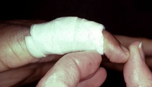
Figure 2: A PIP joint extension immobilization orthosis that allows mobilization of the DIP joint into flexion.
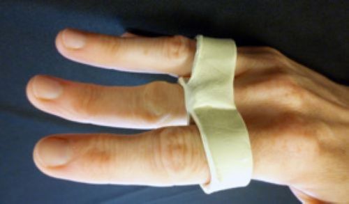
Figure 3: An RMO may be used to treat a boutonniere deformity or a PIP joint flexion contracture.
• After approximately 6 weeks, if full extension is present (See Figure 4.) the patient begins gentle active flexion out of the protective orthosis.
• Once active PIP joint motion is initiated, a relative motion orthosis (RMO) (See Figure 3.) to block MP joint hyperextension may help the patient maintain full PIP joint extension during the day.
• Additionally, blocking the DIP joint in some flexion with a small orthosis increases the lateral band tension, encouraging the lateral bands to move dorsally at the PIP joint, improving PIP joint extension. (See Figure 5.)
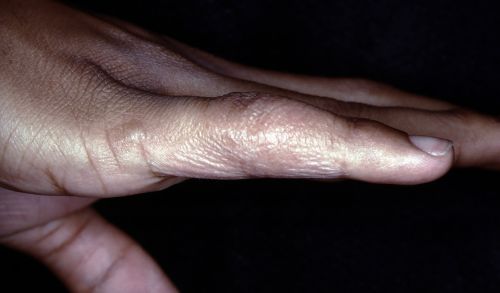
Figure 4: Full active PIP joint extension following immobilization in extension. Note that dorsal thickening gives an illusion of less than full extension.
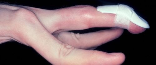
Figure 5: A DIP joint orthosis blocking extension but allowing flexion will assist PIP joint extension.
PIP Joint Flexion Contracture / Pseudo Boutonniere Deformity
A PIP joint immobilization orthosis would worsen symptoms. Instead, focus acute treatment on restoring PIP joint mobility through active motion and controlled stretching. If the PIP joint flexion contracture persists, a relative motion orthosis (RMO) (See Figure 3) can be used to transmit the power of the extrinsic extensors into the dorsal apparatus for better PIP joint extension. The RMO is worn full time for both exercise and activities.
Treatment Decisions
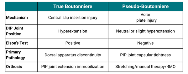
Misidentifying the deformity can lead to prolonged stiffness, delayed recovery, and unnecessary frustration for both clinician and patient. The chart below shows the basic differences between a true boutonniere and a pseudo-boutonniere (flexion contracture).
Conclusion
A boutonniere like posture does not always mean a true boutonniere deformity. By understanding the mechanism of injury, using Elson's test, and considering the timeline, you can confidently differentiate between a central slip insertion injury and a volar plate/capsular joint contracture. This clarity ensures your orthotic and therapeutic interventions truly support recovery—not hinder it.
Download Clinical Pearl No. 97, Boutonniere Basics, July 2026
© BraceLab; 2026 all rights reserved
Disclaimer: BraceLab Clinical Pearls are intended to be an informal sharing of practical clinical ideas; not formal evidence-based conclusions of fact.