
The Role of Skin When Addressing Pain

July 2025 No. 91
The Role of Skin When Addressing Pain
by Karol S. Young, OTD, OTR/L, CHT
As therapists, we often treat issues related to the skin. Realizing the difference between the volar and dorsal skin on the hand, we modify strapping and application of pressure accordingly when fitting an orthosis or providing resistive exercises. We are vigilant to protect the skin integrity when providing orthotic fabrication and fitting in older patients. We also spend considerable time focusing on skin scars. For example, a patient with a Dupuytren’s contracture release where the thicker palmar skin and the palmar fascia pathology produce an adherent and tight scar, we focus on scar management that includes massage, compression and the application of gel sheeting. See Figure 1.

Figure 1: Palmar scarring after a palmar fasciotomy.
But a patient who has been immobilized following a distal radius fracture or patient with CMC osteoarthritis has no visible signs of scarring or skin injury. Do we pay attention to the role that the skin plays when evaluating and treating their pain?
I was recently introduced to Taylor and Wolff’s concepts about pain and the superficial sensory system in lateral elbow pain. The proposed framework of thinking in terms of “nerve length and layers” inspired me take a deeper look into including treatment of the skin with my patients.
Skin as the Source of Pain
Skin is our largest organ. It renews itself every 28 days and helps synthesize Vitamin D. The microbiome of our skin plays an important role in protecting us from infection, regulating our immune system, and maintaining skin health. The skin contains about 60,000 miles of blood vessels and the characteristics of one’s skin differs depending on site location, race, sex and age.
Within the layers of the skin are the somatosensory nerve receptors that communicate pain, temperature, and proprioception to the central nervous system. The type of receptor, location and function of each can be seen in the following chart:

Considering the number of sensory receptors within the skin, it is evident how trauma coupled with compression from a cast, or soft tissue changes from a tight muscle can place tension on the skin and thus on its sensory receptors. Movement of the skin and change of tissue tension can produce pain. Since pain receptors are within the skin layers, we should give more thought to treating pain by targeting the somatosensory nerve receptors within the skin.
Interventions to Decrease Pain
Deep Pressure
Have you ever had a patient who is unable to tolerate the light touch from their sleeve on their scar or skin but reports that deep pressure over the same area is soothing? Deep pressure is known to be calming for neurodivergent individuals and for those with anxiety. The use of deep pressure has also been used to treat individuals with chronic pain.
Deep pressure on the skin can be provided through the following:
1. Foam Wedges: Application of foam wedges in between the fingers may assist with joint alignment during active motion and provides deep pressure to the skin. See Figure 2.

Figure 2. Use of foam wedges to provide deep pressure and joint alignment.
2. Chip Clips: I have used chip clips (spring loaded clips for holding a bag of chips closed) successfully when stretching the thumb web space. Using Ms. Taylor’s concepts, I now use them to place or relieve tension on the skin.
To promote skin glide and provide deep pressure for pain relief, experiment with the placement of the chip clip while obtaining your patient’s feedback on pain relief. See Figure 3.

Figure 3. Use of a chip clip to provide skin glide and deep pressure.
3. Sports Tape: Sports tape is commonly used to treat a variety of acute and chronic conditions including orthopedic injuries. Sports tape, such as Kinesiotape™, can also be used to provide deep pressure to the skin. Taping techniques depend on the patient’s diagnosis and presentation. While taping provides deep pressure and skin glide for pain relief, it can also aid in improving joint alignment and decreasing neural tension as seen in the example of application in Taylor and Wolff’s article.
Vibration
The use of localized vibration is known to produce analgesic effects. According to Casale and Hansson, “localized vibration relieves pain not only by acting on the spinal gate, but also at higher levels of the nervous system.” They found that the best pain reducing frequencies are between 100 and 250 Hz (high frequency) as they involve the spinal gate mechanism. Frequency below 100 Hz (low frequency) may not activate the spinal gate mechanism however, but reduces pain by activating C-mechanoreceptors and interferes with pain at the level of the limbic system. Application with a handheld massager can be used to the patient’s tolerance on the skin, along the length of the nerve and further proximal up the nerve path, to aid in modulating pain. See Figure 4.

Figure 4. Use of a mini massager can be used along a nerve pathway.
Immersion Particles
Immersion of a body part into particles has long been used for sensory re-education to treat hyperesthesia and allodynia. This same technique can reduce skin pain. Immersion particles stimulate touch receptors. Exposure to the particles several times a day for 5 to 10 minutes can gradually reduce skin hypersensitivity. Dry lentils and/or popcorn can be used as a cost-effective immersion medium while commercially available plastic pellets are an easy to clean alternative for use in the hand therapy clinic. Because tactile stimulation activates both the ipsilateral and contralateral somatosensory cortex, simultaneous bilateral immersion may be beneficial. See Figure 5.
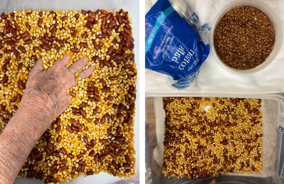
Figure 5. Immersion particles for tactile stimulation.
Pain Within the Skin
Recently, when a painful palmar scar was present in a patient who was returning to production work in the automotive industry, I provided a mini massager and recommended immersion textures to assist with scar desensitization and improve scar pliability. I found these interventions clinically effective but could not specifically explain why the vibration and external stimulation provided pain relief. Thank you, Alison Taylor OTR/L, CHT, CKTI for helping me realize I was also reducing pain within the skin.
Additional Resources Related to Alison Taylor's work:
1. Finkelstein, H: Stenosing tendovaginitis at the radial styloid process. J Bone Joint Surg 12, 1939: 509-40.
2. Harvey EG. Postoperative scar management: strategies for improved outcomes. Clin Dermatol Open Access J. 2024;9(1):000331. doi:10.23880/cdoaj-16000331.
3. Taylor A. Rehab techniques in pain and nerve compression syndromes. Aspetar Sports Med J. 2024;13:23-1.
4. Taylor A. Treating without pain: a new approach to hand therapy. FingerPrint Australian Hand Therapy Association Newsletter. 2024 June;139:25-27.
5. Brutus JP, Taylor A, Barone N, Hagert E. Extensor mechanism dysfunction and hand deformities caused by Dupuytren’s disease: Surgical and rehabilitation perspectives. J Hand Ther. 2025 Feb.
6. Hagert E. Proprioception of the wrist joint: a review of current concepts and possible implications on the rehabilitation of the wrist. J Hand Ther. 2010;23(1):2-17. doi:10.1016/j. jht.2009.07.010. Available from: https://pubmed.ncbi.nlm.nih.gov/19963343/
7. Hagert E. Wrist proprioception—An update on scientific insights and clinical implications. J Hand Ther. 2024;37(2):310-317. doi:10.1016/j.jht.2023.09.004. Available from: https://www.sciencedirect.com/science/article/abs/pii/S0894113023001394
8. Hagert E. Wrist proprioception—An update on scientific insights and clinical implications. J Hand Ther. 2025;38(1):115-122. doi:10.1016/j.jht.2023.09.004. (Republication/feature from 2024 update)
9. Taylor A. Taylor wedges hand pain relief [Internet]. LinkedIn; 2024 Apr 5 [cited 2025 Aug 20]. Available from: https://www.linkedin.com/posts/alison-taylor-0823b5140_walkadifferentdirection-activity-7181891472638582785-iKLJ/
Download Clinical Pearl No. 91, The Role of Skin When Addressing Pain, July 2025
© BraceLab; 2025 all rights reserved
Disclaimer: BraceLab Clinical Pearls are intended to be an informal sharing of practical clinical ideas; not formal evidence-based conclusions of fact.