
Why Can I Not Make A Fist?

May 2025 No. 90
Why Can I Not Make A Fist?
by Karol S. Young, OTD, OTR/L, CHT
Distal radius fractures are one of the most common diagnoses we see in hand therapy; we recently had four referrals for this diagnosis in one day! One patient had been treated with cast immobilization, while the other three underwent open reduction and internal fixation (ORIF) of the fracture. Interestingly, they all had one thing in common. With no passive joint limitations, pain, or significant finger edema, they were still unable to touch their fingertips to their palm when attempting to fully flex their fingers. In frustration they asked: “Why can I not make a tight fist?”
The Immobilization Phenomenon
The intrinsic and extrinsic hand muscles work synergistically to create normal finger motion. After injury, joint stiffness, edema, and healing tissue can disrupt this complex movement system. However, in a hand where none of these conditions are visibly evident in the fingers, how do you explain to your patients the inability to make a fist after wrist immobilization? I call this presentation “the immobilization phenomenon.” See Figure 1.

Figure 1: The inability to make a fist after an injury to the wrist.
To accomplish composite finger flexion, three things must automatically occur:
1. When the extrinsic flexor muscles contract to flex the fingers, the wrist must be able to support the hand with the wrist in extension.
2. The extrinsic flexor digitorum profundus (FDP) muscle must initiate finger flexion at the distal interphalangeal joint (DIP) with a trailing cascade of flexion at the proximal interphalangeal (PIP) and then the metacarpophalangeal joint (MP). See Video 1
3. The interosseous muscles must have enough elasticity to allow the fingers to fully flex.
Video 1: Click to watch video of normal joint motion.
Immobilization after injury or surgery often creates a period of limited total active motion of the fingers, resulting in adaptive shortening of the interosseous muscles (much more so than the lumbrical muscles). The tight interosseous muscles encourage maladaptive motor patterns to develop, with the most common being loss of the active FDP to initiate finger flexion. In this circumstance, finger flexion is initiated at the MP joints.
Common causes of the immobilization phenomenon may include:
• Immobilization of the wrist in a splint/cast that is too far distal to allow full finger flexion.
• Apprehension or fear of movement while in the splint/cast.
• Lack of instruction in proper active finger range of motion.
• Poor or weak wrist control that does not allow the normal tenodesis pattern of wrist extension during finger flexion.
How to Identify the Immobilization Phenomenon
The best way to identify the immobilization phenomenon is to observe your patient’s active movement. This allows you to identify maladaptive motor patterns as well as the wrist position during movement. You should also manually access interosseous muscle tightness.
If the immobilization phenomenon is present, it can be corrected using the treatment techniques described below.
Tools for Recruiting the FDP and Elongating the Intrinsic Muscles
A quick and easy way to facilitate active interphalangeal finger flexion is to apply an MP blocking orthosis. See Figure 2. With the MP joints blocked in extension, active motion of the FDP muscle is redirected to initiate interphalangeal joint flexion rather than allowing the tight interosseous muscles to initiate active flexion at the MP joint.

Figure 2. MP blocking orthosis to encourage active IP joint flexion.

In this position the interosseous and the lumbrical muscles are maximally elongated during active finger flexion. Dots are added to the MP blocking orthosis and the tip of the patient’s fingers. The patient is asked to “touch the dots.” Striving to touch the dots together encourages active MP joint extension which is essential to the elongation (stretch) of both the interosseous and the lumbrical muscles. It is important to remember that the interosseous muscles are “shortened” more readily than the lumbrical muscles and that active IP joint flexion with MP joint extension will elongate both muscles, but passive stretching will only elongate the interosseous muscles.
Another type of orthosis may be fabricated with the MP joints extended and the IP joints in full flexion. The patient is instructed to perform the active motion of tucking the digits into the orthosis. See Figure 3. Once the patient is able to tuck their fingers into the orthosis, then they are instructed to actively extend at the MP joint as much as possible. See Video 2.


Figure 3: Orthosis used to tuck fingers into MP extension and IP flexion.
Video 2: Click to watch video of active MP extension within the orthosis.
Active exercises may also include MP extension with IP flexion over a yoga block or other stable surface and the use of this position to perform resistive exercises into exercise putty. See Figures 4, 5, & Video 3. These active exercises with blocking/resistance provide proprioceptive feedback, helping normalize active movement after immobilization.
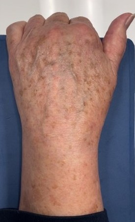

Figure 4: Active hook exercises over a yoga block.
Video 3: Click to watch video of active exercise over yoga block.

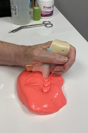
Figure 5: Using putty for resistance while maintaining the active hook position.
While the immobilization phenomenon is often seen in those who have sustained a distal radius fracture, it readily occurs with other diagnoses when the hand/wrist has been immobilized. We prefer to detect maladaptive motor patterns early and begin these exercises in addition to any others needed for the specific diagnosis.
How do you address the immobilization phenomenon in your clinic?
Download Clinical Pearl No. 90, Why Can I Not Make A Fist?, May 2025
© BraceLab; 2025 all rights reserved
Disclaimer: BraceLab Clinical Pearls are intended to be an informal sharing of practical clinical ideas; not formal evidence-based conclusions of fact.