
Treatment After Dupuytren's Fasciectomy...Not Always Simple

September 2025 No. 92
Treatment After Dupuytren's Fasciectomy...Not Always Simple
by Karol S. Young, OTD, OTR/L, CHT
Dupuytren’s disease is the most common genetic connective tissue disorder, and therefore commonly seen in our hand therapy patient population. Following fasciectomy, we care for post operative wounds, work to regain/maintain joint range of motion, and manage the large scar resulting from the extensive dissection required of a palmar fasciectomy. See Figure 1. While these treatments are familiar to hand therapists, comorbidities and joint deformities caused by the Dupuytren’s disease may require special attention. Let’s consider the familiar, but often not so simple post operative care of our patients following palmar fasciectomy.

Figure 1: Post operative fasciectomy of the ring and little fingers.
The Longstanding PIP Joint Contracture
Long standing Dupuytren’s contracture often creates a severe flexion contracture of the little finger proximal interphalangeal (PIP) joint, which may, on occasion, appear as boutonnière deformity.
Patients often wait to seek treatment until they can no longer get their hand into their pocket or cup their hand to wash their face. Unfortunately, delayed intervention promotes continuing tightening of the palmar fascia, often drawing the PIP joint of the involved digit/s into severe flexion. See Figure 2. A longstanding PIP joint contracture changes the balance of the dorsal apparatus of the affected finger, creating adaptive elongation of the dorsal fibers and ultimately perhaps also volar migration of the lateral band, resulting in a boutonnière deformity. See Figure 3.

Figure 2: Dupuytren's disease creating flexion of the little finger joints.

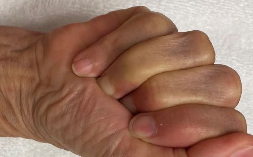
Figure 3: Severe PIP joint contracture resulting in boutonnière deformity of the little finger.
Although the patient and surgeon may wish to regain full PIP joint extension, complete resolution of a long-standing flexion contracture/ boutonnière deformity may not be a realistic goal. When the dorsal apparatus anatomy is altered by adaptive elongation, it loses its mechanical ability to fully extend the PIP joint.
Therefore, when treating a surgically released PIP joint and surrounding fascia, the goal of functional motion rather than full joint extension often produces a more satisfying outcome. Attempts at aggressive correction of the PIP flexion contracture/boutonnière deformity after surgical release often leads to increased pain, swelling, and hand stiffness. Instead, consider the following:
When fabricating a post operative PIP joint extension orthosis:
• Provide only slight passive tension for PIP joint extension.
• Avoid transmitting pressure to the distal interphalangeal (DIP) joint as increased passive DIP joint hyperextension only reinforces the boutonnière posture.
Avoid aggressive passive stretching to the PIP joint. I block the metacarpophalangeal (MP) joint of the little finger from full extension to actively redirect all the muscle forces toward PIP joint extension and increase active distal finger extension. The use of a relative motion orthosis to block MP joint hyperextension may be useful during the daytime to provide the MP joint extension block.
The carpometacarpal joint of the little finger is a mobile joint, thus stabilizing it may help redirect the active motion more distally.
Osteoarthritis, Trigger Finger, and Carpal Tunnel Syndrome
Co-morbidities such as hand osteoarthritis, trigger finger, and/or carpal tunnel syndrome may complicate recovery, as any of these underlying conditions can be exacerbated by the surgery. Following fasciectomy, the body’s natural response to surgery is inflammation. This normal inflammation may, however, trigger increased symptoms of the underlying osteoarthritis or carpal tunnel syndrome. Repetitive stretching and exercises may also aggravate preexisting hand synovitis, resulting in triggering of the finger/s. If concurrent treatment of these conditions is needed, consider the following:
1. Proactively screen for pre-existing conditions at your initial evaluation and modify your treatment plan from its initiation, to avoid exacerbating symptoms.
2. Consider comorbidities when progressing treatment. This may require modifying exercises to avoid repetitive gripping and other positions that exacerbate pre-existing conditions or slowly adding resistance to exercises.
3. Provide specific treatment for these conditions at the onset of symptoms so as to limit their effect on the healing tissues.
Extrinsic and Intrinsic Extensor Balance
Dupuytren’s contracture almost always creates a flexion position of the MP joint of the involved finger. Although prolonged flexion of the MP joint may not create an MP joint contracture, it does change the extrinsic and intrinsic muscle balance that controls both MP and interphalangeal (IP) joint motion.
Prolonged MP joint flexion creates adaptive changes of the extrinsic extensor muscle and the tendon over the MP joint in an elongated position, decreasing its mechanical effectiveness. Conversely, prolonged MP joint flexion also creates interosseous muscle tightness by allowing the interosseous muscles to adaptively shorten. To address the extrinsic and intrinsic extensor imbalance, we recommend:
1. Specifically evaluate for interosseous muscle tightness.
i. Interosseous Muscle Tightness Testing
ii. Nuances of Interosseous Muscle Tightness Testing
2. Provide an active stretch to the interosseous muscles, with active MP joint extension and IP joint flexion with focus on maintaining maximum MP extension as the IP joints flex. If the patient cannot achieve this actively, a removable exercise orthosis to hold the MP joints in extension and allow full active IP joint flexion is recommended.
3. Isolate and strengthen extrinsic extensor muscles by actively extending the MP joints while actively holding the IP joints flexed.
Post operative therapy protocols following Dupuytren’s fasciectomy recommend wound care, passive stretching, active and passive range of motion exercises, scar management, edema control, and progressive strengthening. While these common treatment approaches appear straight forward and suggest a goal of regaining full range of joint motion, experience suggests delayed surgical intervention and comorbidities may make full range of motion an impossible goal. Therefore, post-surgical care following Dupuytren’s fasciectomy requires focused evaluation and implementation of nuanced treatment; it is not always that simple.
© BraceLab; 2025 all rights reserved
Disclaimer: BraceLab Clinical Pearls are intended to be an informal sharing of practical clinical ideas; not formal evidence-based conclusions of fact.